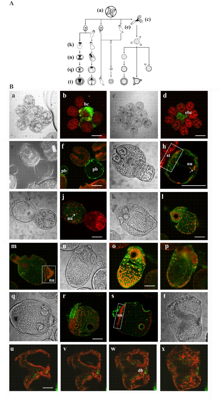
Fig 6. Immunolocalization of Eg-AMPKα during the in vitro de-differentiation process of protoscoleces to microcysts.
(A) Diagrammatic representation of the different mechanisms involved in cyst development during in vitro culture. Image reconstructed using the figure published by Rogan and Richard (1986) [29]. This image is similar but not identical to the original image, and is therefore for illustrative purposes only. (B) Transmission and confocal microscopy of a brood capsule (a,b), an everted brood capsule (c,d), protoscoleces with posterior bladders (e,f), developing cysts from posterior bladders (g-j), vesicularized protoscoleces (k-p) and pre-microcysts developed from vesicularized protoscoleces (q-x). bc: brood capsule; ebc: everted brood capsule; pb: posterior bladder; st: stalk; nu: nucleus; dh: disrupted rostellar hook; broken-lined box indicates stalk portion; solid-lined boxes indicate distribution of nuclei in vesicularized protoscoleces. Bars indicate 200 μm (b and d) and 100 μm (f, h, j, l, o, r and u).

