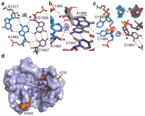
Figure 2.
Solvation at the ribosomal subunit interface and in the nascent peptide exit tunnel. (a–c) Water molecules that bridge the 16S (blue) and 23S (grey) rRNA in bridge B3 of ribosome I. 2′-hydroxyls involved in the water interactions are marked with asterisks. The location of bridge B3 is indicated in the inset. The feature enhanced maps are contoured at 2.5 standard deviations from the mean. (d) Solvation at the entrance of the nascent peptide exit tunnel. Labeled residues of 23S rRNA are shown in orange. The feature enhanced map is contoured at 2.0 standard deviations from the mean.

