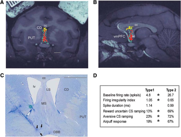
Figure 3.
Locations and firing properties of two groups of reward uncertainty-sensitive neurons in the septal area. A, B, Their locations are shown on MR images: left, coronal; right, parasagittal. Yellow represents Type 1 neurons in the anterodorsal septal region (n = 31). Red represents Type 2 neurons in the mBF (n = 39). Light green represents other recorded neurons (n = 300). C, Electrolytic marking lesions (black arrows) made at the locations of Type 2 neurons in the mBF of Monkey H. They were made along two mediolaterally adjacent electrode tracks. Along the lateral track, we recorded four Type 2 neurons between the two marking lesions. D, A table of response properties of Type 1 and 2 neurons recorded during Experiment 1. Asterisks indicate significant difference between Type 1 and Type 2 neurons (baseline firing rate and irregularity index; Wilcoxon rank sum test, p < 0.05; proportions of neurons displaying significant CSs ramping or airpuff responses; see Materials and Methods; χ2 test, p < 0.05). A–C, Gray lines indicate 5 mm scale bars. A, C, The coronal sections are located ∼1.5 mm anterior to the center of the anterior commissure (ac). cc, Corpus callosum; CD, caudate nucleus; DBB, diagonal band of Broca; lv, lateral ventricle; LS, lateral septum; MS, medial septum; PUT, putamen; vmPFC, ventromedial prefrontal cortex.

