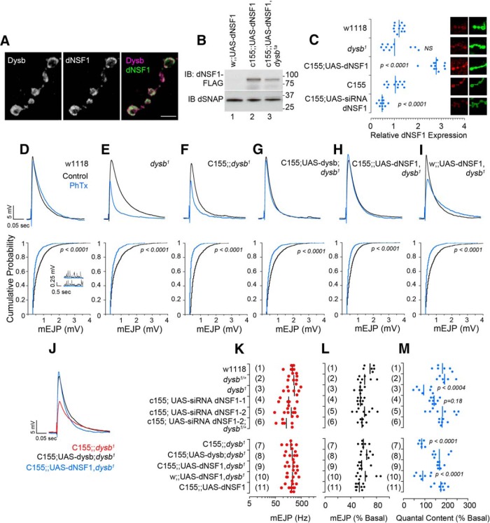
Figure 6.
NSF presynaptically rescues dysbindin synaptic homeostasis defect. A, Confocal microscopy of Drosophila neuromuscular junction stained with antibodies against dNSF1 and GFP to detect functional recombinant neuronal expressed Venus-Dysbindin. Scale bar, 5 μm. B, immunoblot demonstrating the expression of the UAS-dNSF1 transgene in animals with or without the dysb1a mutation. dSNAP was used as a loading control. C, Confocal microscopy of boutons stained with HRP as a control and dNSF1. Fluorescence was normalized per bouton to HRP and to wild-type genotype. D–J, Representative EJP and mEJP traces without PhTx-433 (black) and following PhTx incubation (blue). Representative mEJP traces are shown only for wild-type w1118 animals. J, Overlay of EJP traces after PhTx presented in F–H. K, No differences across genotypes in mEJP frequency in the absence of PhTx. L, No differences across genotypes in mEJP amplitude after PhTx. M, Quantal content across different genotypes. Dots represent individual animals; n > 6 for all genotypes and all conditions. The p values were obtained with one-way ANOVA followed by Bonferroni's all-pair comparisons.

