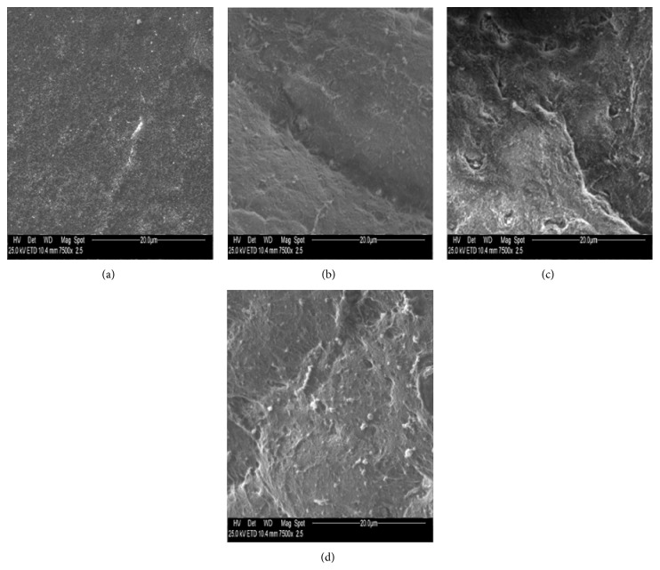
Figure 5.
Photography obtained by electron microscopy (magnification ×7500) of microvilli of cornea rabbit eye before (a, b) and after (c, d) UV irradiation. Corneal epithelium treated with gel formulation without rMnSOD (a, c) and with gel formulation containing rMnSOD 2 μg/mL (b, d). (a) A lot of microvilli widespread on whole surface. (b) A groove as intercellular space, microvilli, and microbumps. (c) A cellular split as intercellular space with dryness and numerous wounds, small microbumps, and few microvilli residues. (d) Microvilli and a few small microbumps. There is no appreciable deterioration of epithelium after exposure to UV radiation.

