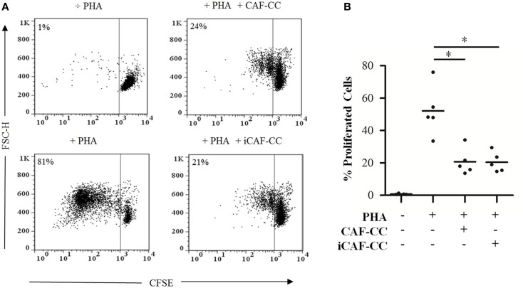
Figure 1.
T-cell proliferation assays: immunosuppressive effects elicited by irradiated and control CAFs during coculture. T-cell proliferation assays were performed using CellTrace™ CFSE labeled human PBMCs activated with 1 μg/ml PHA. Cells were cocultured with irradiated (iCAF-CC) or non-irradiated CAFs (CAF-CC) at a ratio of 1:100 CAF:PBMC and allowed to proliferate for 5 days. Assays were performed with CAFs isolated from five randomly selected donors. T-cell proliferation was determined by measuring CFSE fluorescence intensity by flow cytometry after gating the lymphocyte population by forward and side scatter. (A) Representative flow cytometry dot plots showing percentage of CFSElow-labeled T-cells. One out of five representative experiments is shown. (B) Graph shows rate of T-cell proliferation determined by loss of CFSE fluorescence. Data points are representative for five different CAFs donors with bars representing means from triplicate experiments. Student’s t-test value (*p < 0.05).

