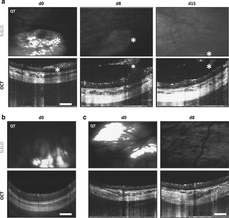
Figure 2.
Detection of trauma, PPC-graft misplacement, and graft loss using bimodal (OCT–fcSLO) imaging. (a) Bimodal imaging showing epiretinal and vitreal graft clouding (asterisks) at day 0 (d0) indicating the presence of a full thickness retinal tear, while at day 8 (d8) and day 15 (d15) these aggregates are composed of mainly host cells; Qtracker (QT) loaded PPCs appear white on fcSLO images. Results are representative images (n=4, recipients: 12–15). (b) Misplacement of graft in the choroid (n=3, recipients: 4–6). (c) QT signal loss indicating graft loss from host site after 8 or 15 days posttransplant (n=8, recipients: 4, 5, 9–11, and 13–15). (a–c) Scale bars are 250 μm.

