Figure 3.
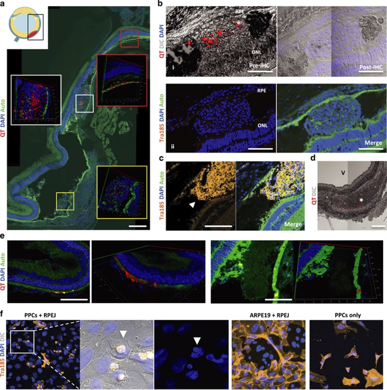
End point Histology of PPC transplants. (a) Representative images of a subretinal bleb containing heterogeneous distribution of Qtracker (QT) graft label at 15 days posttransplant. Nuclei label (DAPI); auto-fluorescence (Auto). (b) (i) Subretinal bleb containing QT exhibiting signal loss after immunohistochemistry (IHC) processing; differential interference contrast (DIC). (ii) Human-specific labeling (Tra185 antibody) did not detect any PPC graft at day 15. (c) Tra185-labeled subretinal PPC-graft (arrowhead) at day 0 posttransplant. (d) Misplaced graft—trace amounts of QT signal in the choroid (asterisk); vitreous (V). (e) Host RPE uptake of QT was found at day 15 posttransplant. (f) Co-cultures of PPCs with rat RPE (RPEJ cell line) showing characteristic phagocytosis of PPCs, while these phagocytosis events were not observed in human (ARPE19 cell line) and rat RPE co-cultures, nor was similar morphology exhibited when cultured alone (PPCs only). (a–e) Scale bars are 250 μm.

