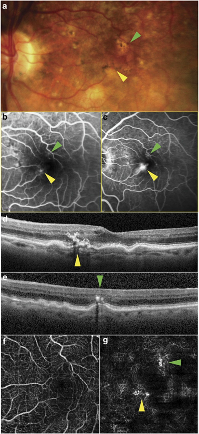
Figure 1.
Multimodal imaging of the left eye of patient 1 showing two type 3 NV lesions bordering the foveal avascular zone. Yellow arrows: high-flow lesion; green arrows: second, low-flow lesion. (a) Color photograph; (b, c) mid- and late-venous phases of fluorescein angiogram; (d, e) SD-OCT through high- and low-flow lesions, respectively; (f, g) en face OCT-A segments of superficial and deep capillary plexus, respectively.

