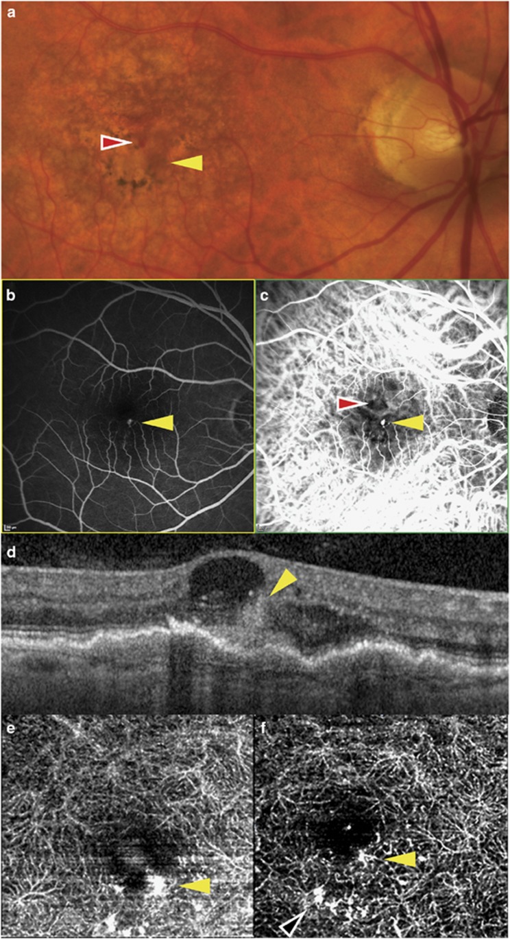
Figure 2.
Multimodal imaging of the right eye of patient 2 showing a type 3 NV lesion (yellow arrow) at the inferior border of the foveal avascular zone, and accompanying intraretinal hemorrhage that is adjacent and superior (red arrow). (a) Color photograph; (b) fluorescein angiogram; (c) indocyanine green angiogram; (d) SD-OCT showing a hyper-reflective intraretinal lesion communicating with a localized pigment epithelial detachment at its outer aspect and an adjacent hemorrhage within a cystic space at its inner aspect; (e) baseline OCT angiogram; (f) OCT angiogram after two intravitreal treatments. Supranormal flow is seen in a region of the capillary plexus inferior to the type 3 lesion (black arrow).

