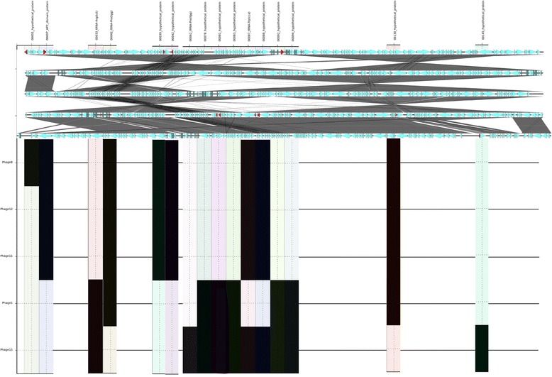
Figure 4.

SeqFindR and Easyfig image combined representing the accessory gene content of group 1. Genomes of each phage in group 1 are represented by the Easyfig image showing linear visualisation of the genome and coding regions represented by arrows, accessory genes are coloured red. The order of phage genomes in the linear visualisation and the accessory content blocking is 8, 12, 11, 1 and 15 and was chosen based on similarity clustering in SeqFindR. Hits for the accessory genes in each genome are represented in labelled columns in the SeqFindR image underneath each accessory gene.
