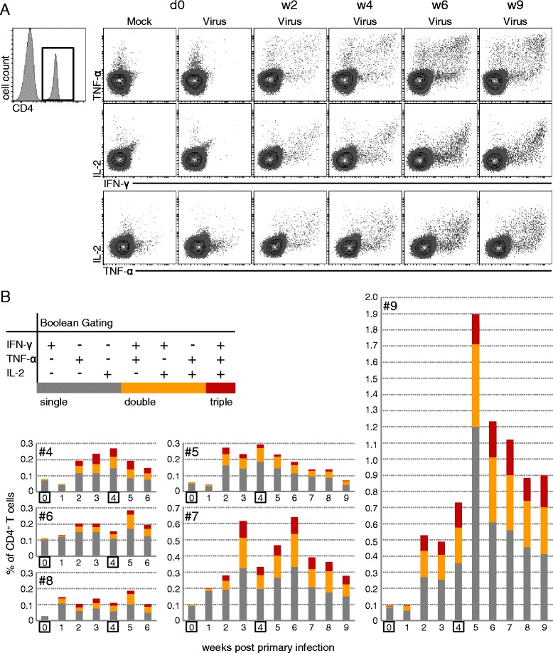
Figure 4.

Kinetics of FLUAVsw-specific IFN-γ-/TNF-α-/IL-2-producing CD4 + T cells. Intracellular cytokine staining of defrosted PBMCs was performed following overnight in vitro restimulation with FLUAVsw (infection strain, MOI = 0.1; 18 h). Mock-incubated cultures served as negative controls. (A) CD4+ T cells were gated and analyzed for production of IFN-γ, TNF-α and IL-2. Contour plots show combinations of cytokines for selected time points following FLUAVsw infection. Exemplary data of animal #9 is shown. (B) Boolean gating was applied in order to identify single-, double-, and triple-cytokine-producing CD4+ T cells. Single- (dark grey), double- (orange) and triple- (red) cytokine-producing cells are shown as percent of total CD4+ T cells for all six infected animals in the time course following FLUAVsw infection.
