
Figure 2. Bcl-2 phosphorylation. (A).
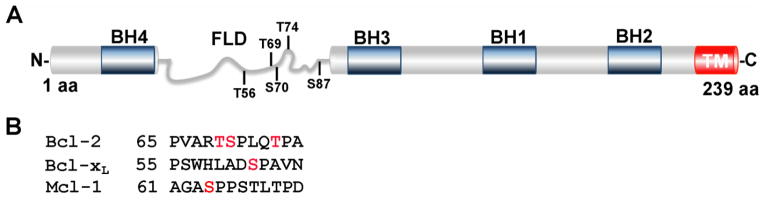
Schematic representation of the Bcl-2 protein with BH domains boxed in blue and flexible loop domain (FLD) labeled in gray. Residues T56, T69, S70, T74 and S87 are located within the FLD and selectively targeted for phosphorylation. S70 (red) is phosphorylated by CDK1 in vivo, inducing a conformation change of the FLD and enhancing the anti-apoptotic function of Bcl-2 [54]. (B). Residues S62 and S64 (labeled in red) within the FLD of Bcl-xL and Mcl-1, respectively, are targeted for phosphorylation by CDK1 [137, 154]. TM, transmembrane domain. (Adapted from [54, 154]).