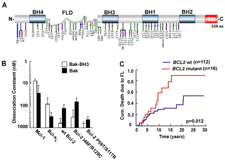
Figure 3. Distribution of Bcl-2 amino acid substitutions in FL. (A).
Schematic representation of the Bcl-2 protein with BH domains boxed in blue and flexible loop domain (FLD) labeled in gray. Somatic variants detected in FL derived from 2 independent cohorts (black and green bars) are distributed throughout the Bcl-2 protein. Color-coded symbols depict distinct types of alterations, with purple for synonymous, white for nonsynonymous with no charge introduction, red for nonsynonymous with negative charge introduction, and blue for nonsynonymous with positive charge introduction. TM, transmembrane domain. (Adapted from Correia et al. [175].) (B). Affinities of Bak BH3 peptide and Bak protein for Bcl-2 variants derived from lymphoid cell lines. (Adapted from Dai et al. [103].) † indicates undetectable binding. (C). Kaplan Meier plots showing impact of BCL2 mutations on death due to lymphoma in a FL cohort (n=128). (Adapted from Correia et al. [175].)

