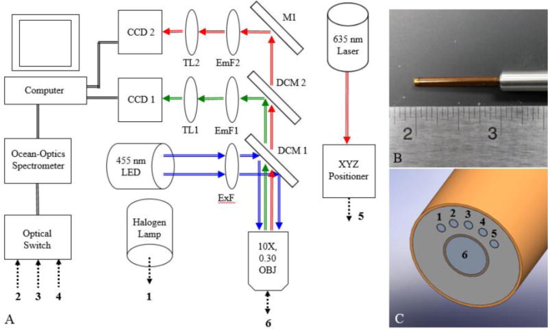
Figure 1.

A representation of the diffuse reflectance and spectroscopic microendoscope (DRSME) showing (A) an instrumentation schematic listing all major components, including numbers (1–6) that indicate the specific fiber number at the proximal end of the fiber-optic probe, (B) an image of the custom-designed fiber-optic probe with a ruler for scale, and (C) a close-up SolidWorks representation of the probe tip with numbers (1–6) that indicate the specific fiber number at the distal end of the fiber-optic probe.
