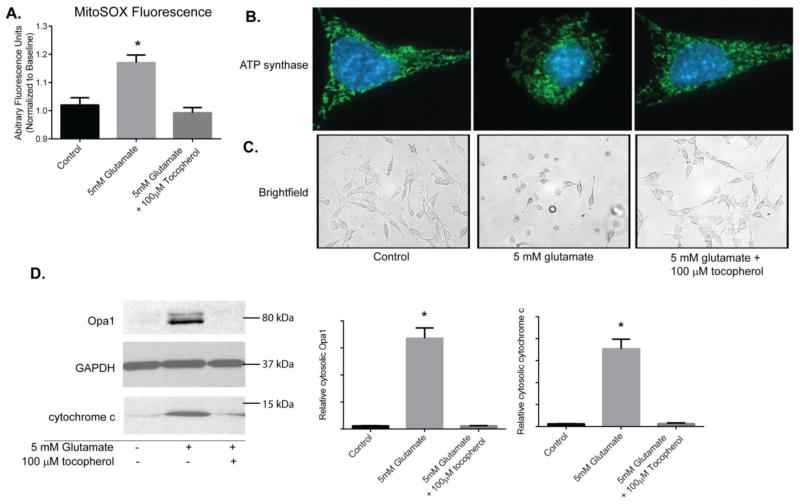
Figure 4. Tocopherol attenuates Opa1 and cytochrome c release following glutamate treatment.
Mitochondrial ROS was measured overtime using MitoSox. 100μM tocopherol prevented ROS accumulation in mitochondria following 5mM glutamate exposure in comparison to glutamate exposed cells (p<0.05; A). Tocopherol was also effective at preserving mitochondrial morphology as observed by immunofluorescence against ATP Synthase (green; B) and maintaining gross morphology of HT22 cells (C). HT22 cells were exposed to 5 mM glutamate alone or 5 mM glutamate with 100μM tocopherol for 10 hours. Cells were fractionated into cytosolic and mitochondrial/heavy membrane fractions. Proteins were resolved on SDS-PAGE and membranes probed for Opa1 and cytochrome c levels. The potent antioxidant tocopherol attenuated both Opa1 and cytochrome c release into the cytosol to levels seen in controls (p<0.05; D).

