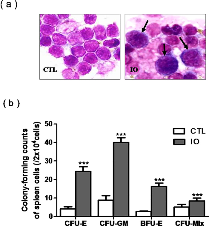
Figure 5. Iron overload led to extramedullary hematopoiesis.

(a) Spleen cells were exposed to Wright's staining (x 1,000). (b) Hematopoietic colony-forming counts (CFU-E, BFU-E, CFU-GM and CFU-mix) from spleen cells are shown as the means ± SE of three independent experiments. N = 6. ***P < 0.001 vs. CTL.
