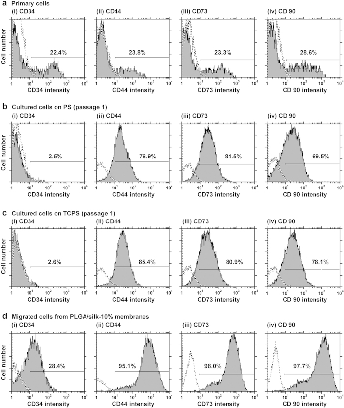
Figure 2. Purity of hADSCs isolated using the hybrid-membrane migration method.
As analyzed by flow cytometry, the expression of CD34 (i) and MSC markers (CD44 [ii], CD73 [iii], and CD90 [iv]) on the primary adipose tissue cells (SVF) (a), first-passage SVF cells grown in untreated PS (b) and TCPS (c) dishes, and the cells that migrated out from PLGA/silk-10% membranes that were subsequently cultured for 15 days after SVF was permeated through the membranes (d). The dotted lines indicate cells labeled with the isotype controls.

