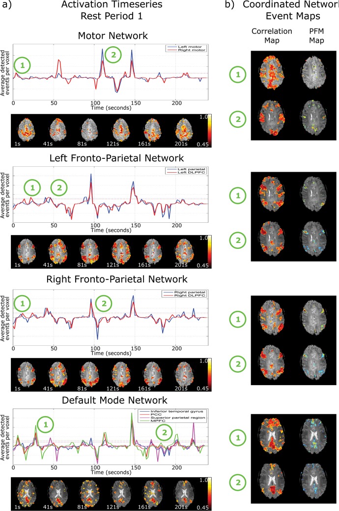
Fig 1. a) Nodal activation timeseries for the motor network, left and right fronto-parietal network and default mode network in rest period 1 from the motor data, for a single subject.
The solid lines show the average number of voxels within the node defined as active by PFM at each time point, and the dotted lines depict one standard deviation from baseline. The correlation maps (below the activation timeseries) are shown for 30s time windows at 40s intervals, each window starting at the time indicated. These highlight the dynamic nature and changing structure of networks b) correlation maps at the time of a coordinated network event that show strong network structure and their corresponding paradigm free mapping activation map depicting the voxels that showed an event at this time.

