
Figure 5.
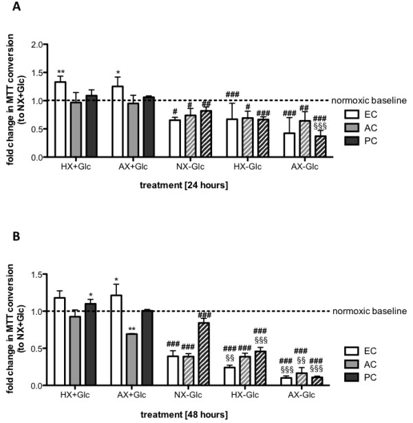
Hypoxic/ischemic exposure differentially modulates metabolic activity of EC and perivascular cells. Viability of primary ECs, ACs and PCs was measured by MTT conversion after exposure to NX, HX and AX in presence (solid bars) and absence (hatched bars) of glucose for 24 h (A) and 48 h (B). Data was normalized to normoxic baseline (NX + Glc). N = 3-5. **P < 0.01, ***P < 0.001; 1-way ANOVA compared to normoxia + glucose. §§ P < 0.01, §§§ P < 0.001; 1-way ANOVA compared to normoxia-glucose. # P < 0.05, ## P < 0.01, ### P < 0.001; 2-way ANOVA comparing + to - glucose. EC: primary endothelial cell, AC: astrocyte, PC: pericyte, NX: normoxia, HX: hypoxia, AX: near anoxia, +Glc: with glucose, -Glc: without glucose.