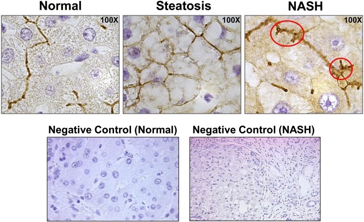
Fig. 3.
Hepatic MRP2 localization in patients with NASH. Immunohistochemistry was used to detect and visualize MRP2 protein localization within normal, steatosis, and NASH liver. The red circles indicate regions of perturbed localization of MRP2 on the canalicular membrane. Negative controls (performed without primary antibody) are included to demonstrate positive membrane staining. All images were taken at 100× magnification and are representative images of multiple immunohistochemical sample analyses.

