Figure 2.

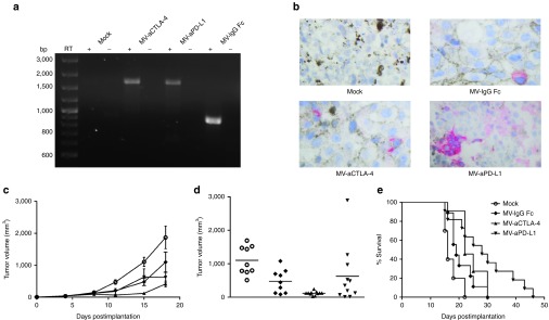
Immunovirotherapy in immunocompetent melanoma model. 1 × 10⁶ B16-CD20 cells were implanted subcutaneously into the flank of C57BL/6 mice. When tumors reached an average volume of 40 mm3, animals were subject to mock treatment (carrier fluid; n = 10) or intratumoral injection of 2 × 10⁶ viral particles of MV-IgG Fc (n = 9), MV-aCTLA-4 (n = 11), or MV-aPD-L1 (n = 11) on 5 consecutive days. (a) Tumors were explanted 24 hours after the last treatment. RNA was extracted and RT-PCR was performed with primers specific for aCTLA-4, aPD-L1, and IgG Fc, respectively. RT- indicates controls without reverse transcriptase. (b) Immunohistochemistry of explanted tumors with an antibody specific for the IgG constant region. Magnification: 500-fold. (c) Tumor volumes were determined every third day. Mean tumor volumes of mock-treated mice and mice treated with the indicated viruses are shown. Error bars represent standard error of the mean. (d) Distribution of tumor volumes for each group on day 15 postimplantation. Dots represent tumor volumes of individual mice. (e) Kaplan–Meier survival analysis.
