
Figure 5.
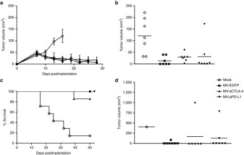
Oncolytic efficacy in human melanoma xenografts. 5 × 10⁶ Mel888 cells were implanted subcutaneously into the flank of NOD/SCID mice. When tumors reached an average volume of 40 mm3, animals were subject to mock treatment (carrier fluid; n = 7) or intratumoral injection of 2 × 10⁶ viral particles of MV-EGFP, MV-aCTLA-4, or MV-aPD-L1 (n = 7 per group) on 5 consecutive days. (a) Tumor volumes were determined every third day. Mean tumor volumes of mock-treated mice and mice treated with the indicated viruses are shown. Error bars represent standard error of the mean. (b) Distribution of tumor volumes for each group on day 16 postimplantation. Dots represent tumor volumes of individual mice. (c) Kaplan–Meier survival analysis. (d) Distribution of tumor volumes for each group on day 50 postimplantation. Dots represent tumor volumes of individual mice.