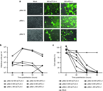
Figure 6.

Infection of primary melanoma. Short-term cultures were prepared from melanoma biopsies and inoculated with recombinant Measles virus (MV) encoding aCTLA-4 or aPD-L1 and EGFP as a reporter gene. (a) Images were taken 48 hours p.i. (MOI 1). Scale bars: 100 µm. Low-passage cells from primary melanoma specimens were infected with MV-aCTLA-4 and MV-aPD-L1. (b) After infection at MOI 3, progeny particles were determined at designated time points. (c) At designated time points after infection at MOI 1, cell viability was determined by XTT assay. Viability of mock-treated cells was defined as 100%. XTT, 2,3-bis-(2-methoxy-4-nitro-5-sulfophenyl)-2H-tetrazolium-5-carboxanilide.
