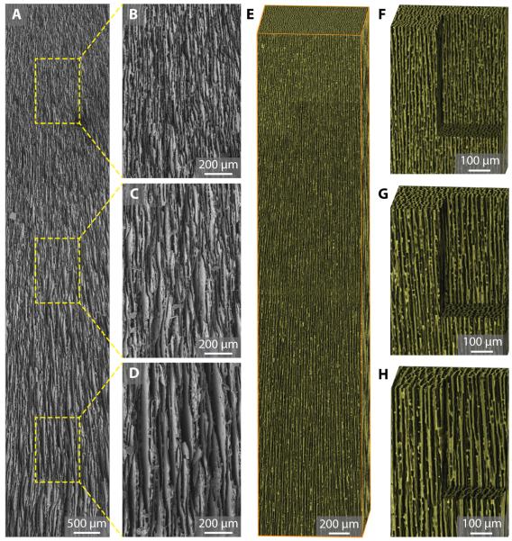
Fig. 2.
Structural characterization of gradient scaffold. (A) Low-magnification SEM image of gradient scaffolds with channel width (λ) increasing from top to bottom. The sample length is around 10 mm. (B-D) High-magnification SEM image at different locations along the channel showing increasing channel width of (B) 4.54 ± 0.88 μm, (C) 8.14 ± 1.24 μm, and (D) 11.8 ± 2.47 μm, respectively. (E) 3-D reconstruction of gradient scaffolds from X-ray micro-tomography data. Bounding box dimension: 1 mm × 1 mm × 5 mm. (F-H) Close-up view of the scaffolds’ inner structure. Scale: 500 μm × 500 μm × 750 μm.

