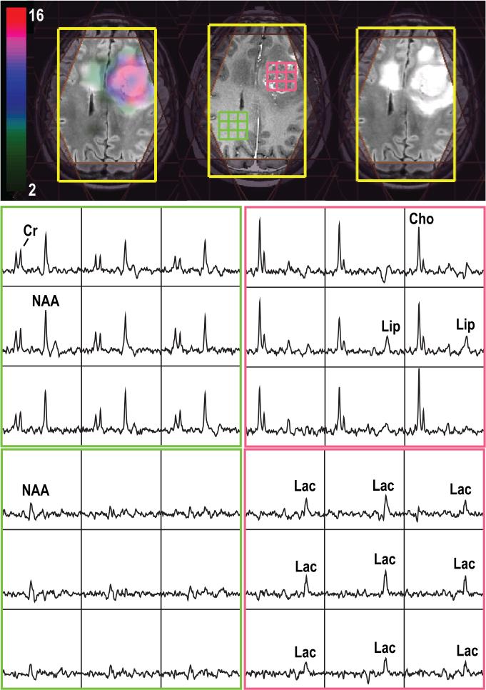
Figure 1.
3D Lac-edited 1H MRSI acquired from a patient with a newly-diagnosed glioblastoma at the time of pre-treatment (TE/TR=144/1500ms, matrix size=18×18×16, elliptical sampling and flyback in S/I, nominal spatial resolution=1cm3, 1 cycle edited on, 1 cycle edited off, total acquisition time=12.96min). Spectral array corresponded to summed (middle row) and difference (last row) of the Lac-edited spectra. The Cho-to-NAA index overlaid on T2-weighted images shows abnormal metabolic lesions.

