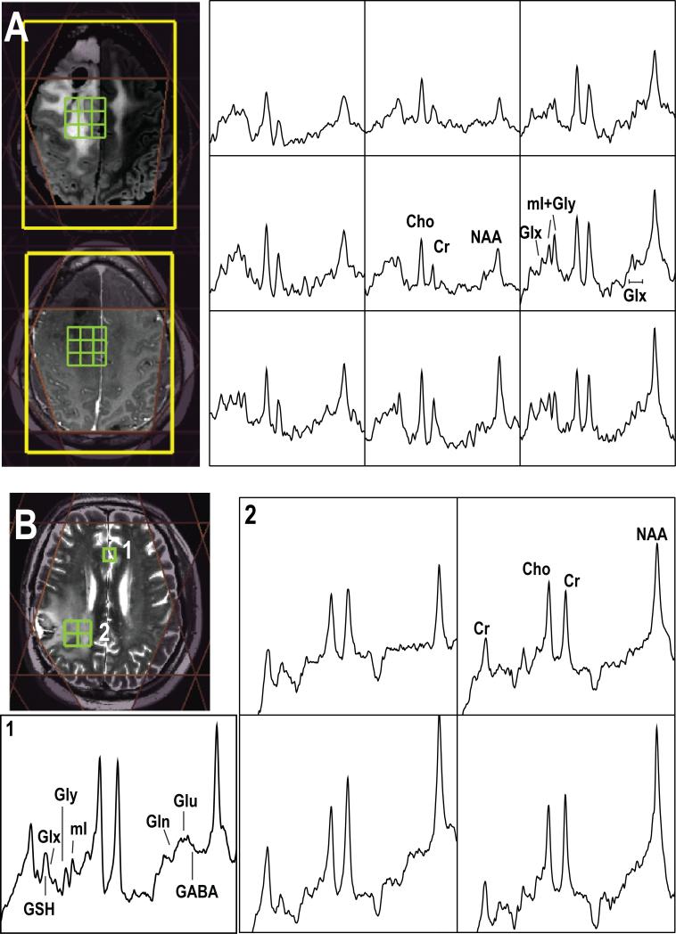
Figure 2.
Examples of short echo 1H MRSI acquired from (A) a patient with grade 2 glioma at 3T (TE/TR=35/1500ms, matrix size=18×18×16, flyback in S/I, nominal spatial resolution=1cm3, total acquisition time=8.1min) and (B) a patient with glioblastoma at 7T (TE/TR=30/2000ms, matrix size=18×22×8, flyback in A/P, nominal spatial resolution=1cm3, total acquisition time=9.6min). Note the baseline has not been removed from the spectra that are shown.

