
Figure 4.
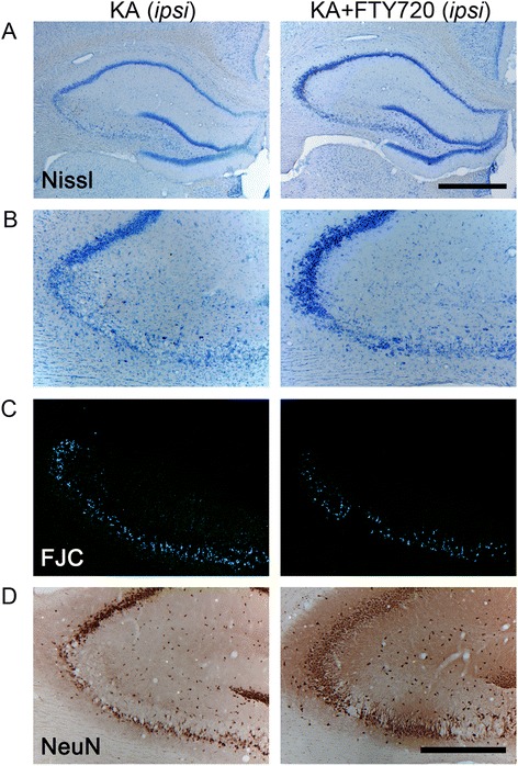
Histological analysis of the protective effect of FTY720 in CA3 region following icv injection of kainic acid (KA). Slices collected 3 days after icv surgery were stained with Nissl (A, B), Fluoro-Jade C (FJC) (C), and anti-NeuN antibody (D). Figure shows representative photomicrographs of bright field (A, B, D) and epifluorescence (C) microscopy images obtained from coronal sections of KA-treated (left panel) and KA + FTY720-treated animal groups (right panel). Only the damaged ipsilateral hemisphere is shown. KA-induced cell death is evident in the CA3 region, as indicated by Nissl’s staining (A, B), lower number of NeuN+ cells (D), and increased density of FJC+ neurons (C). KA + FTY720-treated group (right panel) shows significant attenuated loss of neurons as compared to KA-treated group (left panel). (A) Dorsal ipsilateral hippocampus. Scale bar: 1 mm. (B-D) CA3 region of ipsilateral hippocampus. Scale bar 500 μm.