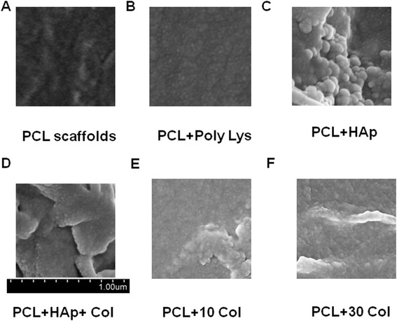
Figure 1.

Representative SEM micrographs of PCL scaffolds after coating. The micrographs show the details of scaffolds after coating. A, PCL scaffolds only; B, PCL scaffolds with poly-lysine coating; C, PCL scaffolds with HAp; D, PCL scaffolds with HAp and 30 mg/ml collagen coating; E, PCL scaffolds with 10 mg/ml collagen coating; F, PCL scaffolds with 30 mg/ml collagen coating.
