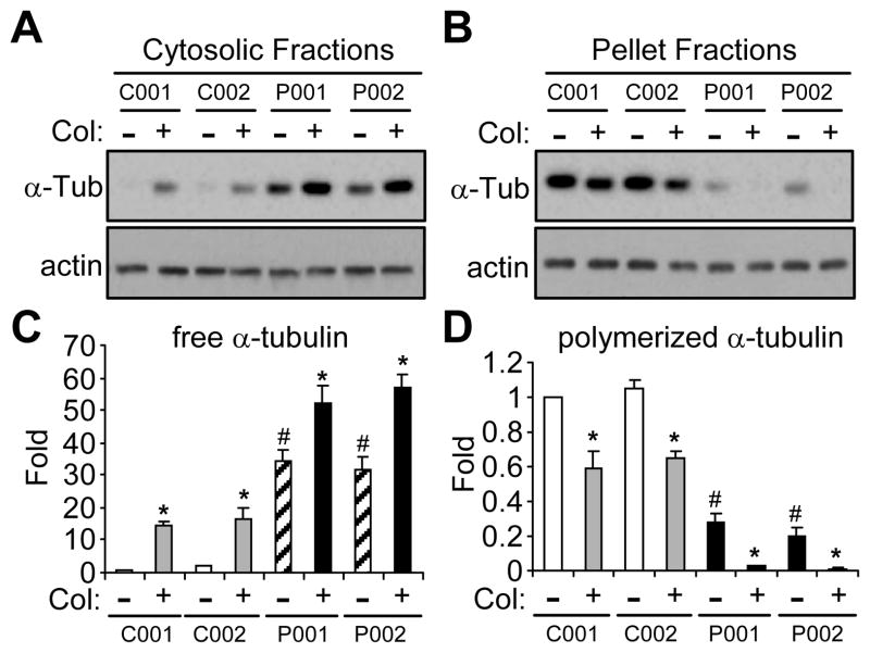
Figure 4.
Microtubule stability in iPSC-derived neurons from normal subjects or PD patients with parkin mutation. (A–D) iPSC-derived neuronal cultures from two normal subjects (C001 and C002) and two PD patients with different parkin mutations (P001 and P002) were treated without or with colchicine (Col, 10 μm for 30 min). Cytosolic (A) or pellet (B) fractions of these cells were blotted with antibodies against α-tubulin or actin. The amount of free tubulin in cytosolic fractions (C) and polymerized tubulin in pellet fractions (D) were quantified by normalizing against the amount of α-tubulin in the first lane. *, p < 0.001, vs. the preceding bar; #, p < 0.001, vs. C001 or C002 without colchicine treatment, n = 3 independent experiments.

