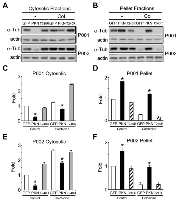
Figure 5.
Microtubule stability in parkin-deficient neurons infected with lentiviruses expressing GFP, wild-type or mutant parkin. (A–F) iPSC-derived neuronal cultures from two PD patients with parkin mutations (P001 and P002) were infected with lentiviruses expressing GFP, wild-type parkin (PKN) or its PD-linked T240R mutant (T240R). After colchicine (Col) treatment (10 μM for 30 min), cytosolic fractions (A) and pellet fractions (B) were blotted with antibodies against α-tubulin or actin. The amount of free tubulin in cytosolic fractions (C, E) and polymerized tubulin in pellet fractions (D, F) were quantified by normalizing against the amount of α-tubulin in the first lane of P001 (C, D) or P002 (E, F), respectively. *, p < 0.001, vs. the preceding bar, n = 3 independent experiments.

