Fig. 4.
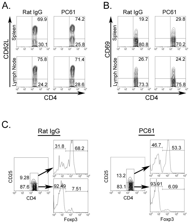
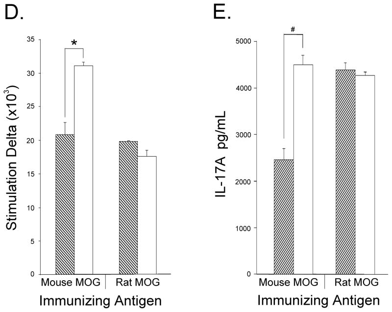
T cells from spleen and LNs of Treg-depleted mice immunized with mouse MOG are hyper-responsive when stimulated with mouse MOG. Mice receiving three i.p. injections of either rat IgG (n=3) or PC61 (n=3) were immunized with mouse MOG and sacrificed on day 20. Spleens or LNs isolated from each group were pooled and CD4+ gated cells were stained for expression of (A) CD62L and (B) CD69. (C) Spleen cells were also tested for anti-CD4, anti-CD25, and anti-Foxp3. (D) Cells isolated from mice immunized with mouse or rat MOG were stimulated with mouse MOG and proliferation was measured as described in Materials and Methods. For (D) data are expressed as stimulation indices and represent one of 2 independent experiments with three mice per group. * p<0.0002, § p<0.003 (E) LN cells from mouse or rat MOG immunized mice were stimulated with mouse MOG protein and supernatants were collected for IL-17A detection by ELISA. Dashed bars- control IgG treated; opened bars- PC61-treated animals


