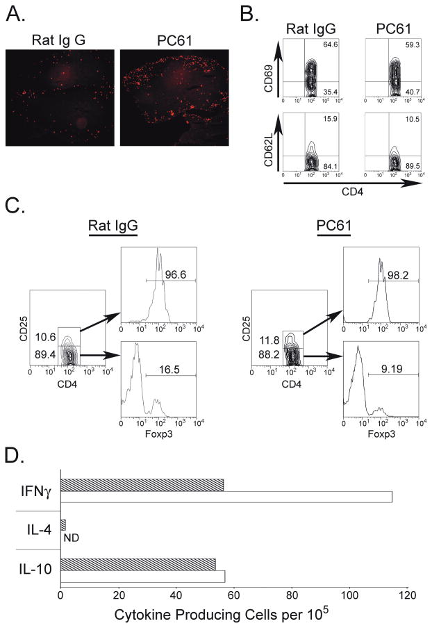
Fig. 5.
Treg depletion results in increased numbers of infiltrating CD4+ cells and increased numbers of IFNγ producing cells in the CNS. Mice treated with either rat IgG (n=3) or PC61 (n=3) were immunized with mouse MOG and sacrificed on day 20. (A) Isolated SC were sectioned and stained for the presence of CD4+ T cells. (B) Mononuclear cells from the CNS were gated on CD4+ cells and stained for the expression of CD62L or CD69. (C) Cells isolated as in (B) were stained with anti-CD4, anti-CD25, and anti-Foxp3. (D) ELISPOT analysis. CNS tissues from each group were pooled together and 105 cells were plated and stimulated with either mouse MOG or MOG35-55. Dashed bars- control IgG treated; open bars- PC61-treated animals. Data are expressed as stimulation indices and represent one of 2 independent experiments with three mice per group.

