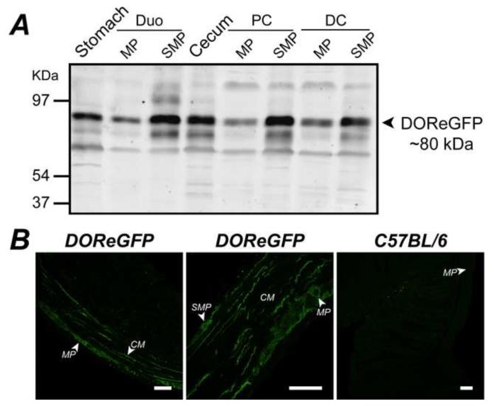
Figure 1. Expression and localization of DOR in the gastrointestinal tract.
A. DOReGFP was detected as ~80 kDa protein in Western blots of stomach, duodenum (Duo), cecum, proximal colon (PC), and distal colon (DC), including muscularis externa/myenteric plexus (MP) and submucosa/submucosal plexus (SMP). B. DOReGFP-IR was localized to neurons of myenteric and submucosal plexuses and to nerve fibers within longitudinal and circular (CM) smooth muscle layers (left and middle panels) of colon. There was no detectable GFP-IR distal colon of wild-type mice (right panel), demonstrating specificity. Scale, 50 μm.

