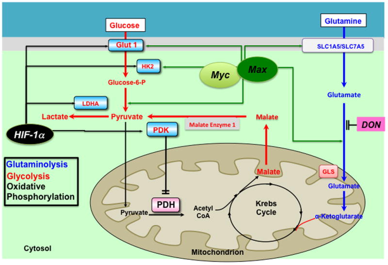
Figure 3.

Proposed mechanism of glutaminolysis in the hypertrophied right ventricle. RV ischemia and capillary rarefaction activate cMyc and its binding partner Max, which increases the expression of the glutamine transporters (SLC 1A5 and 1A7) and augments glutamine uptake. This drives the production of α-ketoglutarate (α-KG). α-KG enters Krebs’ cycle leading to production of malate. Krebs’ cycle-derived malate generates cytosolic pyruvate, which is converted by lactate dehydrogenase A (LDHA) to lactate. In conditions of high glutaminolysis, glucose oxidation is inhibited. DON can inhibit glutaminolysis and restore glucose oxidation. HIF-1α increases the transcription of the some of the same glycolytic mediators as cMyc and Max, notably Glut1 and HK2. Abbreviations: Glut1 = Glucose transporter 1, HK = hexokinase, HIF-1α= Hypoxia inducible factor 1α, LDHA = lactate dehydrogenase A, PDH = Pyruvate dehydrogenase, PDK = Pyruvate dehydrogenase kinase, PFK = phosphofructokinase. DON= 6-diazo-5-oxo-l-norleucine. Reprinted with permission from 21.
