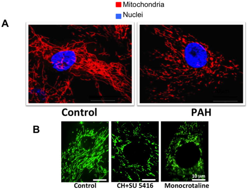
Figure 4.

Mitochondrial fragmentation in Pulmonary Arterial Hypertension (PAH). A. Mitochondria are more fragmented in PAH versus control pulmonary artery smooth muscle cells (PASMCs). Quantification of the mitochondrial fragmentation count reveals a doubling of the number of individual mitochondria in PAH versus control PASMCs. Scale bar = 20 μm. Reprinted with permission from 10. B. Increased mitochondrial fragmentation observed in PASMC of rats with PAH induced by exposure to chronic hypoxia plus the VEGF receptor antagonist, SU5416 (CH+SU 5416) or monocrotaline. Mitochondria were imaged by infection of cells with BacMam virus carrying a mitochondrial-targeted green fluorescent protein transgene. Reprinted with permission from 12.
