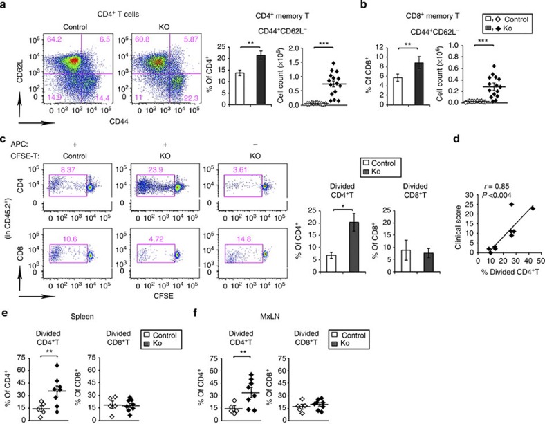
Figure 4. CD4+ T cells from CD11c-Flip-KO mice are autoreactive.
(a,b) pLNs were examined for memory T cells (CD44+CD62L−) in CD4+ (a) and CD8+ (b) T-cell populations at ≥20 weeks in the CD11c-Flip-KO (KO) and control mice. (c) In vitro syngeneic mixed lymphocyte response was employed to identify autoreactive T cells. T cells from the pLNs and brachial LNs of ≥20 week CD11c-Flip-KO mice were CFSE-labelled and represent the responder T cells (3 × 10⁵). The antigen-presenting cells (APCs) are T-cell-depleted spleen cells from CD45.1 mice (3 × 10⁵). After co-culture for 5–7 days, cell division was determined by the dilution of CFSE. The panels on the left are representative flow gating defining the divided cells present in the CD45.2+ CD4+ or CD8+ T-cell populations. The panels on the right are the analysis of KO (n=9) and control mice (n=4). (d) Correlation (Pearson's) between the severity of arthritis in Flip-DC-KO mice just before being killed and the percent of divided CD4+ T cells as defined in c. Identification of autoreactive T cells by adoptive transfer of CFSE-labelled T cells from the (e) spleen and (f) MxLNs of CD11c-Flip-KO or littermate control mice (≥20 weeks, CD45.2+) into CD45.1 recipients. The autoreactive T cells were defined using flow cytometry as defined in c. The spleens and MxLNs from the recipients were examined 8–11 days after transfer. Each data point represents an individual recipient mouse that received T cells from a single donor (*P<0.05, **P<0.01 and ***P<0.001, unpaired two-sided t-test).

