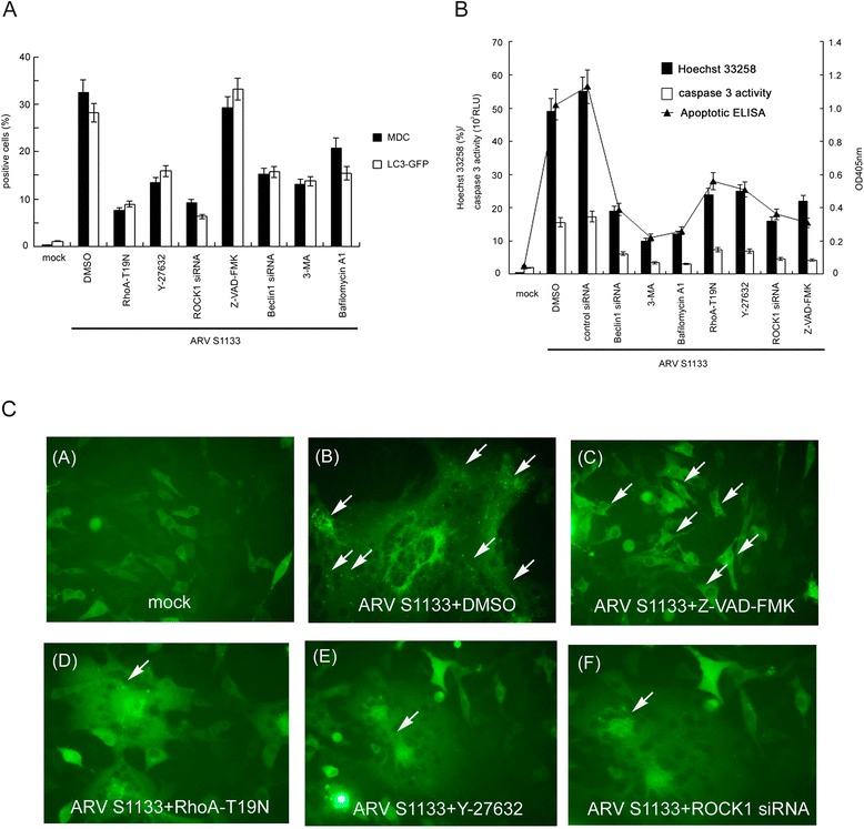
Figure 4.

RhoA and ROCK1 are essential for ARV S1133-induced autophagy, apoptosis, and the conversion of autophagy to apoptosis. (A) Vero cells transfected with RhoA-T19N and incubated for 24 hr, before being transfected with siRNA for 72 hr, pre-treated with 5 μM Y-27632, 10 μM Z-VAD-FMK, 50 μM 3-MA and 0.1 μM Bafilomycin A1 at non-toxic concentrations for 4 hr, and then infected with ARV S1133 at an MOI of 5 for an additional 18-hr incubation. Autophagic vacuoles were stained with MDC, and the percentage of positive cells was calculated in 20 independent fields at a magnification of 200× (black bar). Vero cells transfected with LC3-GFP, together with RhoA-T19N, and incubated for 24 hr, or transfected with siRNA for 48 hr then transfected with LC3-GFP, or pretreated with inhibitor for 4 hr then transfected with LC3-GFP. 24 hr after LC3-GFP transfection, ARV S1133 was added for an additional 18-hr incubation period. GFP-positive cells containing more than 3 dots were counted as positive LC3-GFP cells. The percentage of positive cells was calculated in 20 independent fields at a magnification of 200× (white bar). (B) ARV S1133 infected cells at an MOI of 5 with an incubation period of 36 hr with identical treatment timings to those described in (A). Three apoptosis assays were performed. The left y-axis represents the percentage of Hoechst 33258 positive cells and the caspase-3 activity in relative light units (RLUs). The right y-axis represents the OD 405 nm values from an apoptotic ELISA assay. All experiments were performed three times, each in duplicate. The data are presented as the mean ± SD. (C) Punctate dots of GFP indicating autophagosomes are shown by the white arrows (400x magnification).
