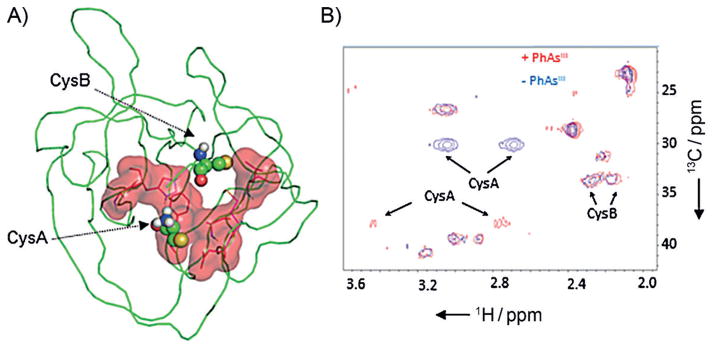
Figure 3.

PhAsIII binding site on Hh located by NMR chemical shift mapping. A) Proposed binding site. Residues in 1H-/15N-labeled HINT domain of Hh whose resonances were suppressed by >80% following the addition of 4 equiv of PhAsIII are displayed in red (sticks), along with the calculated surface area. Catalytic cysteine residues (CysA and B) are shown as ball-and-stick. This figure was generated from PDB ID: 1ATO by using PyMOL (http://www.pymol.org). B) Chemical shift perturbation of CysA in the PhAsIII -bound HINT domain. 13C/1H spectra of the Hh HINT domain enriched with Cys-13C3 at CysA and B, acquired without (blue) and with (red) added PhAsIII.
