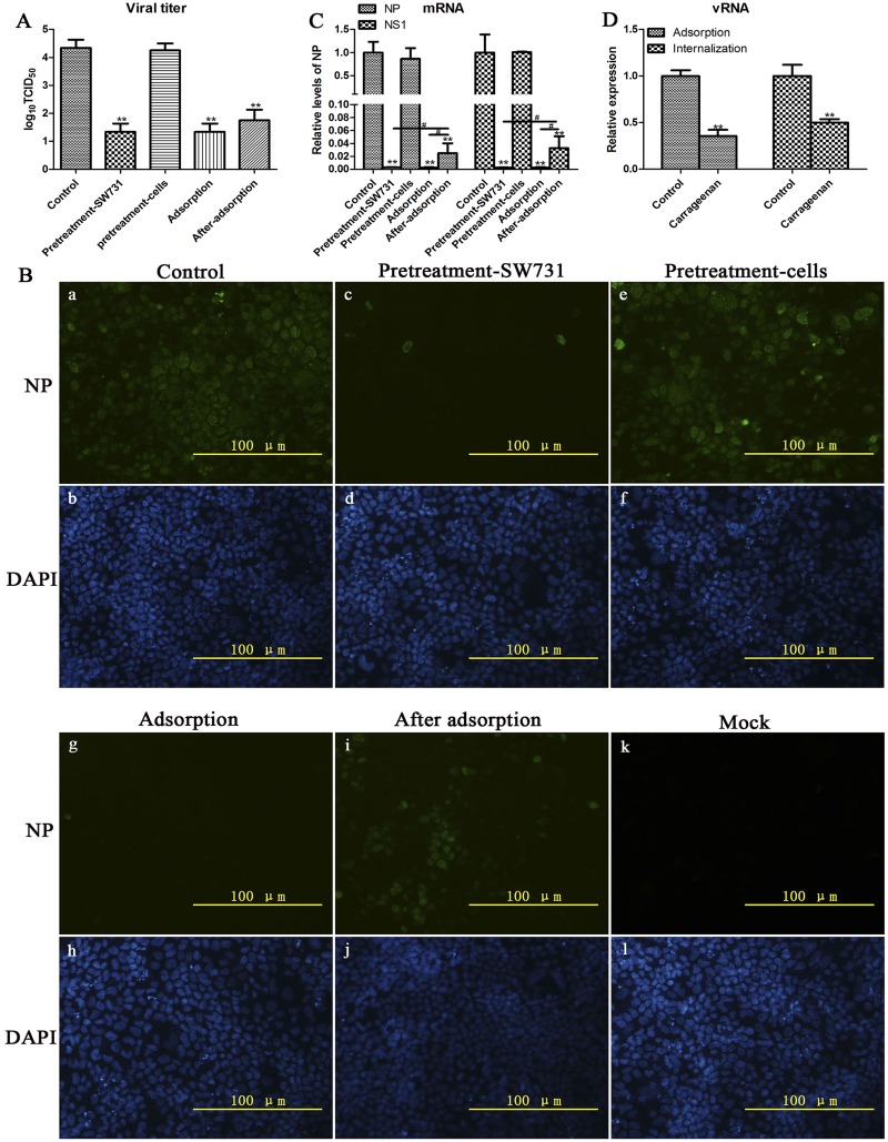
Fig 4. κ-carrageenan-induced inhibition of viral adsorption and internalization.
(A–C) MDCK cells were infected with SW731 (MOI = 3) under 4 different treatment conditions. (i) Pretreatment-SW731: SW731 was mixed with 250 μg/ml of κ-carrageenan at 4°C for 1 h before adsorption. Then the virus/compound mixture was added to cells for 1 h at 4°C, and the media were removed and washed with PBS for three times to remove the compound. The cells were maintained in infective media at 37°C for 24h; (ii) Pretreatment-cells: MDCK cells were treated with 250 μg/ml of κ-carrageenan at 37°C for 1 h before infection. After removing the compound, cells were washed with PBS and incubated with virus for 1 h at 4°C. Then cells were maintained in infective media at 37°C for 24h; (iii) Adsorption: cells were incubated with virus/carrageenan mixture for 1 h of adsorption at 4°C. Then cells were washed and overlaid with infective media at 37°C for 24 h; (iiii) After-adsorption: cells were incubated with virus for 1 h at 4°C. After removal of unabsorbed virus, cells were overlaid with infective media containing 250 μg/ml of κ-carrageenan for 1h at 37°C. Then cells were washed with PBS and maintained in compound free infective media for 24 h. Viral titers were determined in a series of TCID50 assays. The mRNA levels and NP-positive cells were detected using qRT-PCR and immunofluorescence staining, respectively. (D) MDCK cells were infected with SW731 (MOI = 3) by 2 different treatment conditions. (i) Adsorption: MDCK cells were incubated with SW731 in the presence or absence (control) of κ-carrageenan at concentration of 250 μg/ml. One hour later, the media were removed and total RNA was isolated. (ii) Internalization: MDCK cells were incubated with SW731 at 4°C for 1 h. After removal of the inoculation, the cells were overlaid with infective media in the presence or absence of κ-carrageenan at concentration of 250 μg/ml at 37°C for 1 h, after treatment with protease K for 5 min, total RNA was extracted and analyzed using qRT-PCR. Results were analyzed using the independent sample t test. Values are means ± SEM (n = 3). Significance: *P < 0.05 vs. nondrug treated control group; **P < 0.005 vs. nondrug treated control group; # P < 0.05 vs. after adsorption group. Results are representative of two independent experiments.

