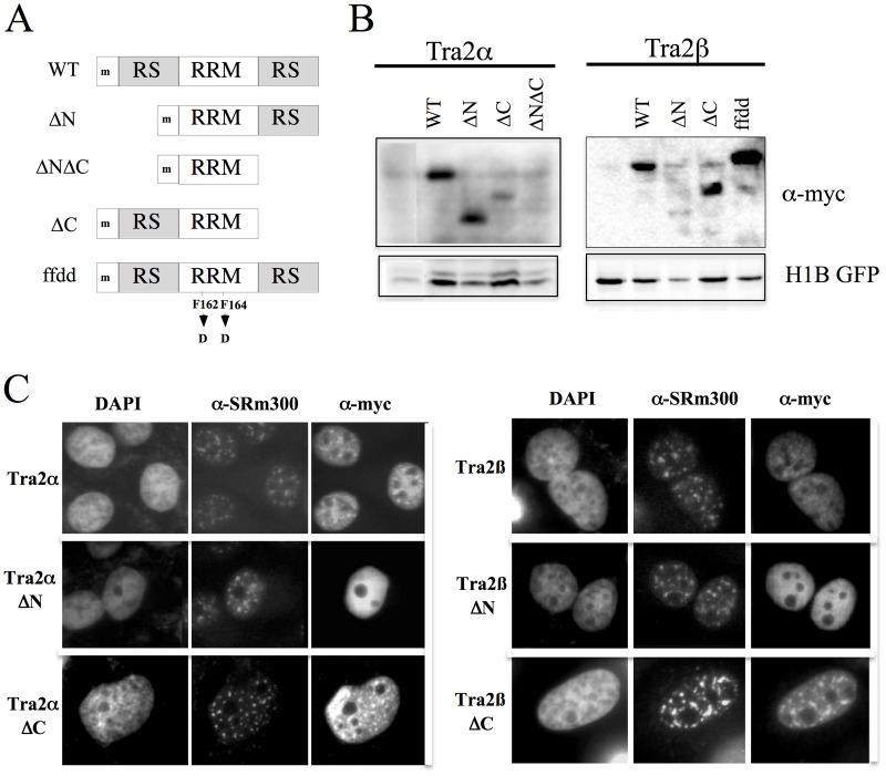
Fig 1. Structure, Expression and Subcellular Distribution of Tra2α/β Mutants.
A, Schematic representation of the panel of deletion mutants created for Tra2α and Tra2β. RRM- RNA recognition motif, RS- arginine-serine rich domain, ‘m’ indicates the presence of an N-terminal myc epitope tag. WT refers to the full length protein. B, To examine expression of the Tra2 variants generated, 293T cells were transfected with the myc-epitope tagged constructs indicated as well a plasmid expressing histone 1B fused to GFP (H1B GFP) to normalize for transfection efficiency. Total cell lysate was prepared and fractionated on SDS-PAGE gels. Blots were subsequently probed with anti-myc antibody. C, HeLa cells were transfected on coverslips with CMVmyc Tra2α and mutants thereof (left panel) or CMVmyc Traβ and mutants thereof (right panel). Samples were processed 48 hours post-transfection for the subcellular localization of transfected Tra2α/β proteins and SRm300 as outlined in “Materials and Methods”. Localization of the proteins was determined by indirect immunofluorescence using α-myc antibody and polyclonal α-SRm300 antibody. Position of nuclei was determined by DAPI staining.

