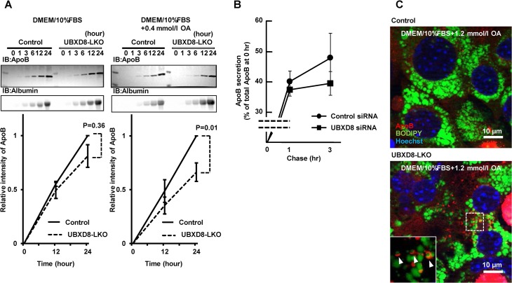
Fig 7. Comparison of cultured cells.
(A) Cultured primary hepatocytes obtained from the UBXD8-LKO mice secreted lower levels of ApoB than cells obtained from the control mice. Hepatocytes obtained from female mice fed a normal diet until 12 wk of age were cultured, and ApoB and albumin secreted into the culture medium were examined by densitometry of Western-blot signals. The albumin signal was used for normalization. The amount of secreted ApoB was lower in hepatocytes obtained from UBXD8-LKO mice than in those obtained from control mice, whether the cells were cultured in the normal medium (DMEM + 10% FBS) (left) or in the medium supplemented with 0.4 mmol/l OA (right). P values were obtained by non-paired Student's t test (n = 3; means ± SEM). (B) HepG2 cells transfected with control or UBXD8 siRNA were pulse-labeled with 35S-methionine and treated with 0.4 mmol/l oleic acid. ApoB immunoprecipitated from the medium at 1 and 3 h was subjected to SDS-PAGE and quantitated by radiography. ApoB secreted in the medium is shown as the ratio relative to total cellular ApoB immediately after pulse-labeling. (C) Primary hepatocytes obtained from UBXD8-LKO mice showed ApoB-crescent structures. The ApoB-crescent was observed as a crescent-shaped ApoB labeling (red) adjacent to LDs (green). Nuclei were stained with Hoechst dye (blue). The ApoB-crescent was present only in cells derived from UBXD8-LKO mice, and not in those obtained from control mice. Cells were cultured in the presence of 1.2 mmol/l OA.

