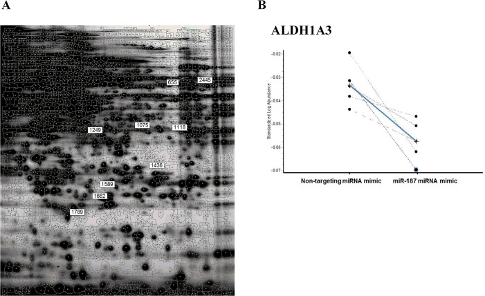
Fig 2. Identification of miR-187 putative targets by 2D-DIGE and LC-MS/MS.
PC-3 cells were transfected either with miRNA mimic negative control or miR-187 miRNA mimic, harvested after 72h, and protein lysates were labeled with Cy3 or Cy5 (miR-187 and control) and Cy2 for the internal standard. A) 2D-DIGE gel image obtained at pH 3–10 and 12,5% SDS-polyacrilamide. The numbers refer to the identification given to the spots differentially expressed. Spot 655 was further identified by LC-MS/MS as ALDH1A3. B) Comparison of the expression of one of the spots (655 or ALDH1A3), in the six gels analyzed, between the cells transfected with miR-187 or with the negative control. The average fold change between the two conditions was -1.06 with a p-value of 0.003. ALDH1A3, aldehyde dehydrogenase family 1 member A3

