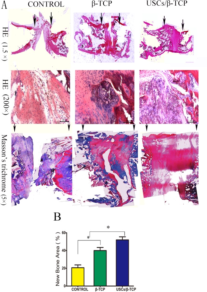
Fig 7. Histological analysis reveals bone formation within empty control, β-TCP and USCs/β-TCP at 12 weeks.

(a) HE and Masson’s trichrome staining showing the repair of bone formation in the critical size femoral defect model. Arrows indicate the edges of host bone. (b) The percentage of the new bone area was calculated from the image of Masson’s Trichrome sections (*P<0.05). Scale bar = 100 μm (HE 200×).
