
Figure 3.
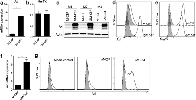
GM-CSF drives Axl expression on macrophages. (a) Axl and (b) MerTK relative mRNA expression in M-CSF- or GM-CSF-differentiated bone marrow-derived macrophages (BMDMs). (c) Western blot analysis of Axl protein expression in M-CSF- or GM-CSF-differentiated BMDMs. M1–M3 represent lysates of cells obtained from individual mice. Expression of (d) Axl and (e) MerTK in M-CSF- or GM-CSF-differentiated BMDMs analyzed by flow cytometry. Specific staining, solid line/open. Isotype control, dotted line/shaded. (f) Relative mRNA expression of Axl in peritoneal macrophages measured after either M-CSF (50 ng ml−1) or GM-CSF (50 ng ml−1) stimulation for 24 h. (g) Flow cytometric analysis of regulation of Axl expression on peritoneal macrophages isolated from naïve mice. Peritoneal macrophages were left untreated or were stimulated for 48 h with M-CSF (50 ng ml−1) or GM-CSF (50 ng ml−1). Specific staining, solid line/open. Isotype control, dotted line/shaded. Data are representative of two independent experiments with three or four mice. Quantitative PCR data are expressed as the mean relative gene expression±s.e.m. of three or four individual mice (a, b, and f). *P<0.05, **P<0.01 vs. corresponding group; unpaired t-test.