
Figure 5.
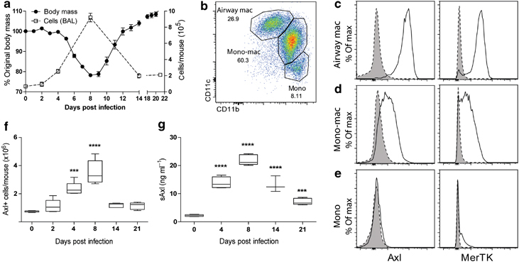
Increase in the numbers of Axl-expressing cells during influenza. (a) Change in body mass of wild-type mice infected with 7.5 p.f.u. influenza (closed symbol) and change in total cell numbers in the bronchoairway lavage (BAL; open symbol), assessed at 0–21 days after infection. (b) Flow cytometric analysis of F4/80-positive cells in the BAL from influenza-infected mice (8 days post infection) by CD11b and CD11c expression. Airway macrophages (airway mac), lung monocyte-macrophages (mono-mac), and lung monocytes (mono) are defined as in Figure 1a. (c–e) Flow cytometric analysis of Axl and MerTK expression on airway macrophages, monocyte-macrophages, and monocytes in the BAL from mice infected with influenza (8 days post infection). Specific staining, solid line/open. Isotype control, dotted line/shaded. (f) Number of Axl-expressing airway macrophages and monocyte-macrophage populations in the total lung during the course of influenza infection (0–21 days post infection). (g) Amount of soluble Axl (sAxl) released into the BAL fluid during the course of influenza infection. Data are representative of two independent experiments with five mice. ***P<0.001, ****P<0.0001 vs. naïve group (day 0); one-way analysis of variance.