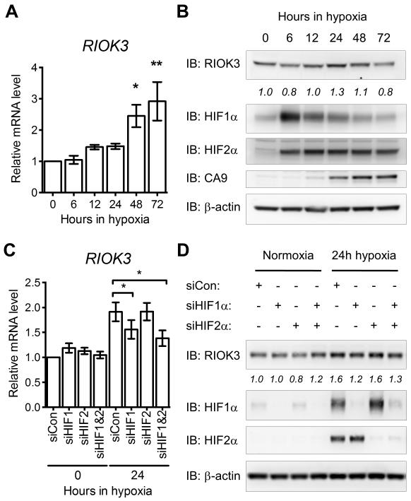
Figure 1.
RIOK3 expression is increased in MDA-MB-231 cells during hypoxia in a HIF1α-dependent manner. (A) Expression of RIOK3 mRNA is increased in MDA-MB-231 cells during exposure to hypoxia (mean ± SEM, n = 3). (B) Expression of RIOK3 protein is transiently increased in MDA-MB-231 cells during exposure to hypoxia. Band density is indicated by italicised numbers below the immunoblot. HIF1α, HIF2α and CA9 immunoblots are presented as hypoxic controls. (C) The hypoxic up-regulation of RIOK3 mRNA in MDA-MB-231 cells is suppressed following transfection with siRNA targeting HIF1α or HIF1α and HIF2α (mean ± SEM, n = 4, repeated measures one-way ANOVA). (D) The hypoxic up-regulation of RIOK3 protein in MDA-MB-231 cells is suppressed following transfection with siRNA targeting HIF1α or HIF1α and HIF2α. Band density is indicated by italicised numbers below the immunoblot.

