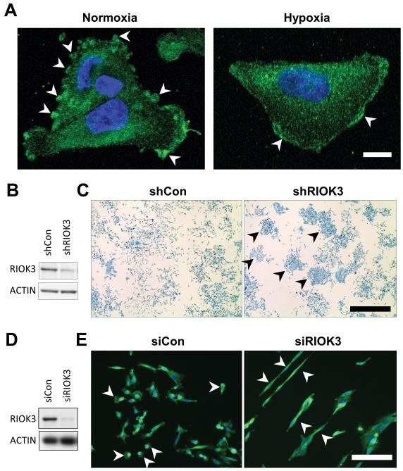
Figure 2.
RIOK3 undergoes subcellular re-localisation in hypoxia and its depletion alters cell morphology. (A) Representative confocal images of migrating MDA-MB-231 cells in normoxia (Nor) or hypoxia (Hyp) stained for RIOK3. Scale bar = 10 μm. (B) Immunoblot demonstrating RIOK3 expression in MDA-MB-231 cells transduced with shCon or shRIOK3 lentivirus. (C) shCon cells grow in a homogeneous, scattered pattern, whereas shRIOK3 cells form colonies after 1 week in culture (arrowheads). Scale bar = 1 mm. (D) Immunoblot demonstrating RIOK3 expression in MDA-MB-231 cells transfected with siCon or siRIOK3. (E) MDA-MB-231 cells immunofluorescently stained for RIOK3. Scale bar = 50 μm.

