Figure 8.
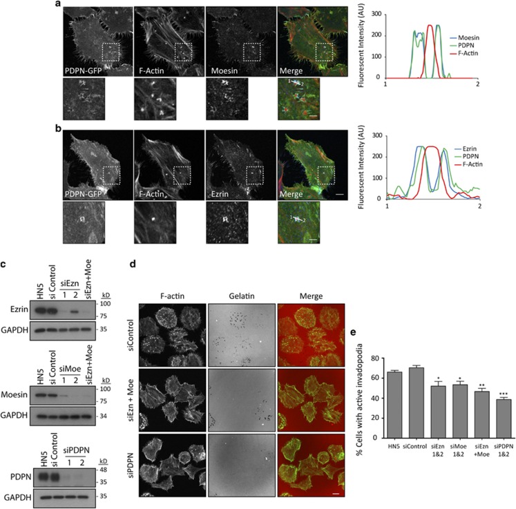
ERM proteins localise at invadopodia adhesion rings and mediate podoplanin ring assembly. (a, b) Specific localisation of moesin (a) and ezrin (b) at invadopodia of HN5 cells cultured on crosslinked gelatin. Note that ezrin and moesin cluster to invadopodia adhesion rings where they colocalise with podoplanin. Graphs indicate fluorescent intensity (in arbitrary units) of each marker over the indicated line scan. Data shown are representative from five invadopodia analysed per condition from three independent experiments. Bars=10 μm (upper panels) and 5 μm (lower panels). (c) Western blot analysis of ezrin, moesin and podoplanin expression in HN5 cells upon specific siRNA treatment. (d) Gelatin-degradation assay (6 h) of HN5 cells treated with ERM and podoplanin siRNAs. Bars=20 μm. (e) Quantification of the gelatin-degradation assay depicted in d. The results shown are the means±s.e.m. of n⩾100 cells for each condition over three independent experiments. ***P<0.0005; **P<0.005; *P<0.05.

