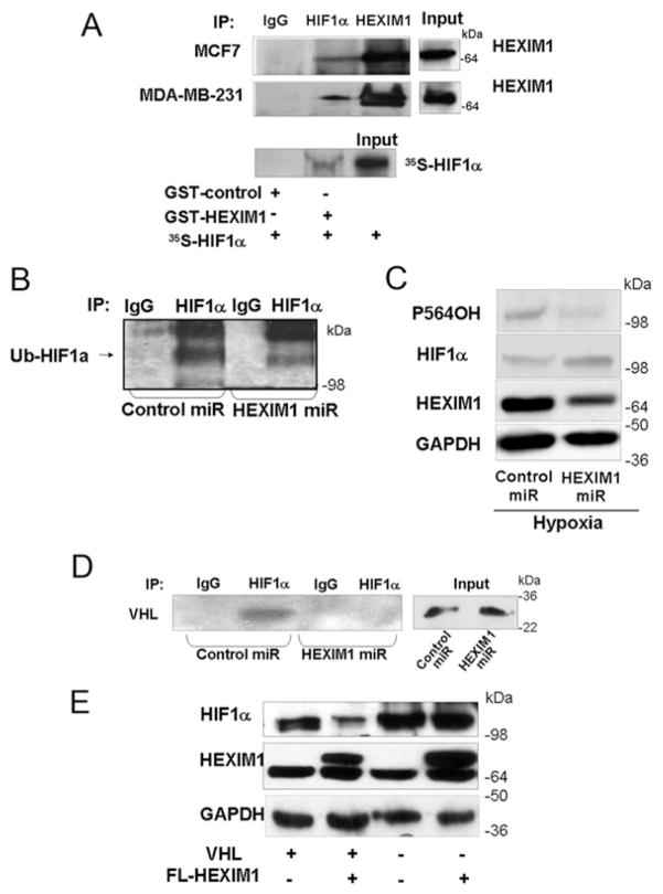
Figure 2. HEXIM1 interacted with HIF-1α and down-regulation of HEXIM1 resulted in decreased levels of hydroxylated and ubiquitinated HIF-1α and attenuated the HIF-1α–pVHL interaction.
(A) Upper panels, MCF7 cells and MDA-MB-231 cells were subjected to hypoxia treatment (8 h). Lysates were immunoprecipitated (IP) using antibodies against HIF-1α or HEXIM1 and analysed for co-immunoprecipitating proteins by Western blotting using HEXIM1 antibody. Normal rabbit immunoglobulin was used as a specificity control. Input lanes represent 25 % of the total protein. MCF7 and MDA-MB-231 panels represent five and three experiments respectively. Lower panel, in vitro translated and [35S]methionine-labelled HIF-1α was incubated with GST or GST–HEXIM1 bound to Sepharose. Input lane represents 10 % of the total volume of in vitro translated product used in each reaction. Panels represent eight experiments. (B) Lysates from control miRNA or HEXIM1 miRNA transfected MCF7 cells were immunoprecipitated using anti-HIF-1α antibody and analysed by Western blotting using the indicated anti-(pan-ubiquitinated HIF-1α) (Ub-HIF1a) antibody. Cells were subjected to hypoxia and MG132 (10 uM) treatments to accumulate ubiquitinated HIF-1α. Panels are representative of three experiments. (C) Western blot analyses of HIF-1α hydroxylated at Pro564 (P564OH) and total HIF-1α in lysates from hypoxia-treated control miRNA or HEXIM1 miRNA transfected MCF7 cells. Panels are representative of six experiments. (D) Control miRNA or HEXIM1 miRNA transfected MCF7 cells were subjected to hypoxia and MG132 (10 uM) treatments. Lysates were immunoprecipitated using anti-HIF-1α antibody and analysed for co-immunoprecipitating proteins by Western blotting using anti-pVHL antibody. Normal rabbit immunoglobulin was used as a specificity control. Input lanes represent 25 % of the total protein. Panels are representative of three experiments. (E) pVHL-deficient RCC4 or wild-type pVHL transfected RCC4 cells were transfected with control vector or expression vector for FLAG (FL)–HEXIM1. The cells were incubated under hypoxia for 8 h as indicated. The expression of HEXIM1, HIF-1α and GAPDH were analysed by Western blot analyses. Panels are representative of four experiments. Molecular masses in kDa are shown next to the Western blots.

